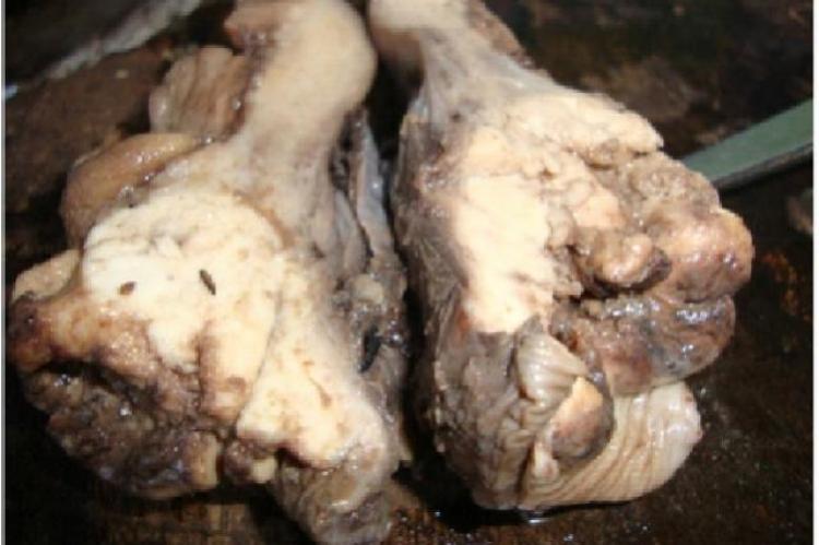
Glassy Cell Carcinoma is a rare malignancy of uterine cervix with aggressive biological behaviour and poor prognosis. Patients usually present with abnormal bleeding or discharge per vaginum of a short duration. It affects comparatively young women of 3rd to 4th decades than other invasive carcinomas of cervix. Histologically it is diagnosed by sheets of large polygonal cells with distinct cell borders, finely granular ground glass like cytoplasm and vesicular nuclei with prominent nucleoli. This is a rare subtype of poorly differentiated adenosquamous carcinoma and proved immune histochemically by positivity to CEA, CK7 and Pan CK with HMWK. But cytologic diagnosis from pap smear can be difficult and can be established by detection of large round to polygonal cells with finely granular cytoplasm and prominent nucleoli. Finding of an inflammatory background is a helpful adjunt to a correct diagnosis. Since the present case was a young patient, surgery followed by chemotherapy was given with a good response. Although rare, such tumours should be kept in mind while dealing with younger patients of suspected carcinoma of cervix. We present a case of glassy cell carcinoma of cervix in a 30 year old female diagnosed in pap smear with clue to cytologic interpretation.
View:
- PDF (1.51 MB)


