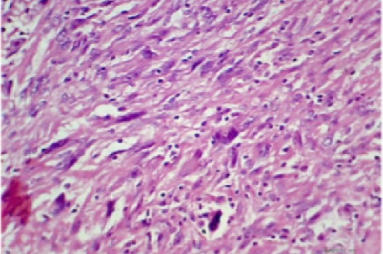
Mature cystic teratoma (MCT) makes up almost 20% of all ovarian neoplasms. They are unilateral in 88% of cases and bilateral in about 10% of cases. Malignant neoplasm is an uncommon event in MCT. It occurs in approximately 2% of cases. Most common malignant change in MCT is Squamous cell carcinoma, followed by carcinoid tumour, adenocarcinoma, malignant melanoma, sarcoma of various types, carcinosarcoma and malignant tumour of neural tissue. In this study we present a case of 38 years old woman presented with complains of pain and lump in the abdomen. She was evaluated clinically and biochemically. Investigation by ultrasound showed a left ovarian cystic mass with mixed echo pattern suggestive of Dermoid cyst with solid area. Her CA 125 was high. She underwent a hysterectomy with bilateral salpingo-oophorectomy for the removal of ovarian mass. On gross as well as on histopathology examination tumor shows features of Dermoid cyst with area showing highly cellular, spindle shaped cells with features of anaplasia and increased abnormal mitotic activity suggestive of poorly differentiated malignant tumor. Immunohistochemical study (IHC) shows strong positivity for cytokeratin confirms poorly differentiated (Sarcomatoid) Carcinoma arising in a mature cystic teratoma of the ovary. This rare type of malignant transformation should be kept in mind when faced with a dermoid cyst, especially
View:
- PDF (867.23 KB)


