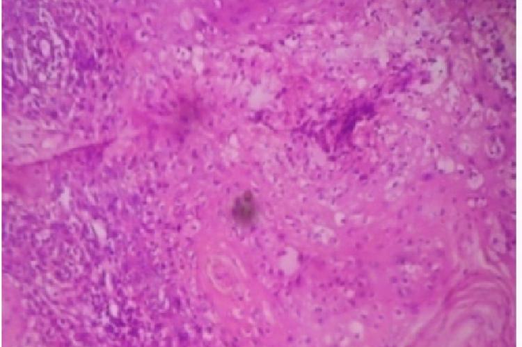
Enlarged lymph nodes are easily accessible for fine needle aspiration cytology (FNAC) and plays major role in diagnosis of primary and secondary malignancies. Correct diagnosis of metastatic tumors by FNAC saves patient from invasive and costly diagnostic procedures and helps the surgeons to formulate therapeutic strategy in treatable primary tumors. Patients presented with palpable lymphadenopathy and suspicious of metastasis referred for cytological evaluation and cytologically diagnosed as metastatic lymphadenopathy from September 2011 to September 2013 were collected. Thorough examination of the patients and detailed clinical history was taken. Multiple smears were studied and clinicocytological correlation was done to find out the primary tumor. Total number of cases studied was 64. The commonest group of lymph node was cervical followed by axillary and inguinal group. Maximum number of cases was in the age group of 50-70 years Male to female ratio was 1.4:1. Nasopharynx was the commonest primary site followed by breast. In 11% cases tongue was primary site and in 5% cases esophagus. Oropharynx and larynx was primary in 2 cases. Primary from lung, parotid and perennial skin was noted in 1 case each. Histopathological follow-up was available in 30 cases. Histopathological correlation of cytologically confirmed primaries showed concordance in 28 cases and 2 cases were discordant FNAC is rapid and safe technique. Thorough clinicocytological evaluation is effective method to detect the primary tumor and act as cost effective diagnostic procedure. Hence this study was done to evaluate the utility of clinicocytological study in diagnosis of primary in metastatic lymphadenopathy.
View:
- PDF (1.39 MB)


